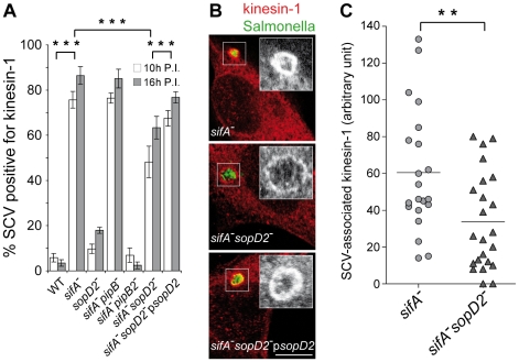
Figure 6. SopD2 influences the retention of kinesin-1 on the bacterial vacuole.
HeLa cells were infected with various GFP-expressing strains fixed and immunostained for kinesin heavy chain and LAMP1 as a SCV membrane marker (not shown). (A) Deletion of sopD2 reduces the percentage of kinesin-1-positive sifA − SCVs. Kinesin-1-positive SCVs were scored at 10 and 16 h p.i.. Values are means ± SD of 3 independent experiments. (B and C) Deletion of sopD2 decreases the amount of kinesin-1 on sifA− SCVs. (B) HeLa cells infected for 16 h were imaged by confocal microscopy for GFP (green) and kinesin-1 (red). Scale bar, 10 µm. (C) Quantitative analysis of confocal images was used to determine the relative staining intensities for kinesin-1 of sifA− and sifA−sopD2− vacuoles. Each point corresponds to the analysis of one SCV. Results of one experiment are shown. This experiment was repeated three times, with comparable results. P-values: ** P<0.01; *** P<0.001.

