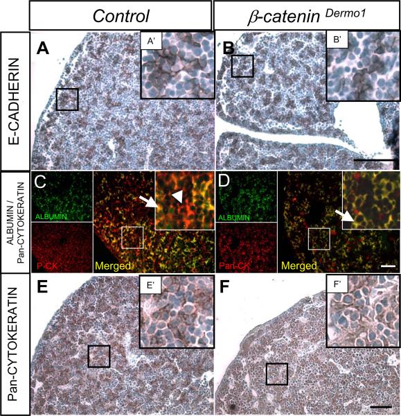
Figure 4.
Immunohistochemical analysis of epithelial markers E14.5 β-CateninDermo1 embryo livers. Staining for E-CADHERIN in the E14.5 β-CateninDermo1 livers (B, B') and littermate control (A, A'). Immunofluorescent staining for ALBUMIN (green), Pan-CYTOKERATIN (red) and merge (arrow, yellow) (C, D). Arrowhead denotes Pan-CYTOKERATIN only positive cells in the control compared to β-CateninDermo1. Immunohistochemical staining for pan-CYTOKERATIN mark bile duct epithelial cells and hepatoblasts (brown) (E, F' and insets E', F'). Bars denote 50 µm

