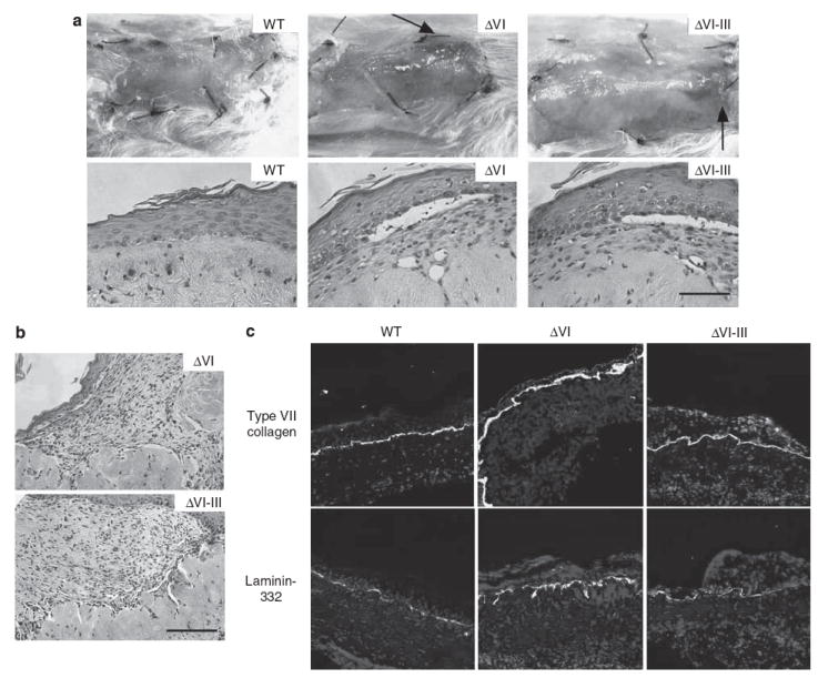
Figure 1. Clinical, histological, and immunofluorescent microscopic analysis of primary human xenografts expressing engineered laminin mutants.

(a) Clinical (upper panels) and microscopic (lower panels) appearance of human skin equivalents 3 weeks after xenografting to severe combined immunodeficiency (SCID) mice. Grafts expressing wild-type (WT) and mutant (ΔVI, ΔVI–III) laminin-332 are shown as indicated (the arrows depict foci of granulation tissue in mutant grafts). (b) Microscopic appearance of granulation tissue arising in mutant laminin xenografts. (c) Laminin-332 and type VII collagen expression in xenografted skin as shown by immunofluorescent microscopy using the indicated antibodies. Bar = 100 μm.
