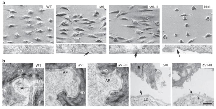
Figure 2. Electron microscopic analysis of primary keratinocytes expressing engineered laminin mutants.

(a) Scanning electron micrscopy (upper panels) and transmission electron microscopy of the cell-culture surface interface (lower panels) of primary human junctional epidermolysis bullosa (JEB) laminin β3 null keratinocytes, retrovirally transduced with WT mutant ΔVI, mutant ΔVI-III, or vector control (Null) cDNA. Arrows in the lower panels point to areas of poor apposition of keratinocyte plasma membrane with the culture surface. (b) Transmission electron microscopic analysis of the indicated intact (left three panels) or separated (right two panels) graft areas. Arrows point to the lamina densa (LM). Abbreviations: AF, anchoring fibril; HD, hemidesmosome. Bar = 1 μm.
