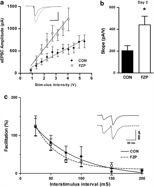
Figure 1.
The slope of the input–output (I/O) relationship, but not paired-pulse facilitation (PPF) was increased in CA1 neurons from 2-day FZP-withdrawn rats. (a) Evoked EPSC (VH=−80 mV) amplitude (pA) was plotted as a function of stimulus intensity (V). Linear regression of pooled I/O relationships showed an ∼2.5-fold increase in the stimulus response relationship in CA1 neurons from FZP-withdrawn (open circles, n=9) compared with control (closed circles, n=7) rats. Inset: Representative traces of eEPSCs elicited at 3.6 V (solid line: CON; broken line: FZP). Scale as in inset in (c). (b) The mean slope of the I/O curve derived from the fits of individual I/O curves generated in neurons from FZP-withdrawn rats (440.0±79.4 pA/V, n=9) was also significantly greater than that from control rats (205.3±44.5 pA/V, n=7). The data are consistent with an increase in CA1 neuron AMPAR synaptic efficacy in FZP-withdrawn rats. Asterisks denote p<0.05. (c) PPF of AMPAR was unchanged in 2-day FZP-withdrawn rats. The amplitude of AMPAR-mediated eEPSCs (VH=−80 mV) after half-maximal stimulation of the Schaffer-collateral pathway. Paired-pulse stimulation was applied and the response was recorded at the interstimulus intervals ranging from 25 to 200 ms in 25 ms increments. Inset: Representative paired EPSC traces recorded in CA1 neurons from control and FZP-withdrawn rats. Paired EPSC amplitudes were calculated as the difference between baseline before the stimulus artifact and EPSC peak. PPF was calculated as (EPSC2−EPSC1)/EPSC1 × 100. Percent facilitation was plotted (CON, closed circles, n=5; FZP: closed circles, n=5) and fit with a single-exponential decay function. No significant differences between groups were found at any interstimulus interval suggesting that glutamate release onto CA1 neurons was unaltered.

