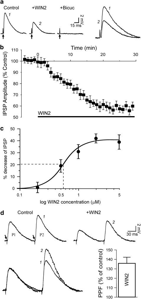
Figure 2.
WIN2 decreases GABAA-mediated IPSPs in CeA neurons at a presynaptic site. (a) Representative recordings showing IPSPs elicited before (control) and during superfusion of 2 μM WIN2 (applied for 25 min) in a CeA neuron. The CB1 ligand diminished the evoked IPSP. The addition of bicuculline completely obliterated the synaptic response, confirming the GABAA nature of the IPSP. Recordings were performed at RMP, −82 mV for this neuron. Stimulus artifact indicated by arrows; traces identified with numbers are magnified and superimposed on the right for comparison. (b) Graph average of IPSP amplitudes over time. WIN2 (application indicated with bar) was added at t=0, and the maximum effect was obtained after 20 min of superfusion. (c) Dose-response curve (logistic fit) of the WIN2 effect. WIN2 decreased IPSPs by a maximum of 41%, with an apparent EC50 of 0.55 μM (dashed line). (d) Upper panel: with the PPF paradigm, WIN2 (2 μM) diminished both IPSPs (P1 and P2) but increased the paired-pulse ratio (P2/P1). Arrows indicate stimulation artifact. Lower left: recordings from panel c superimposed and normalized to P1 to magnify the increase of PPF. For equivalent P1 amplitudes, P2 (identified with numbers) was larger in the presence of WIN2. Lower right: bar graph average depicting the PPF change relative to control condition: 2 μM WIN2 increased PPF by 36%.

