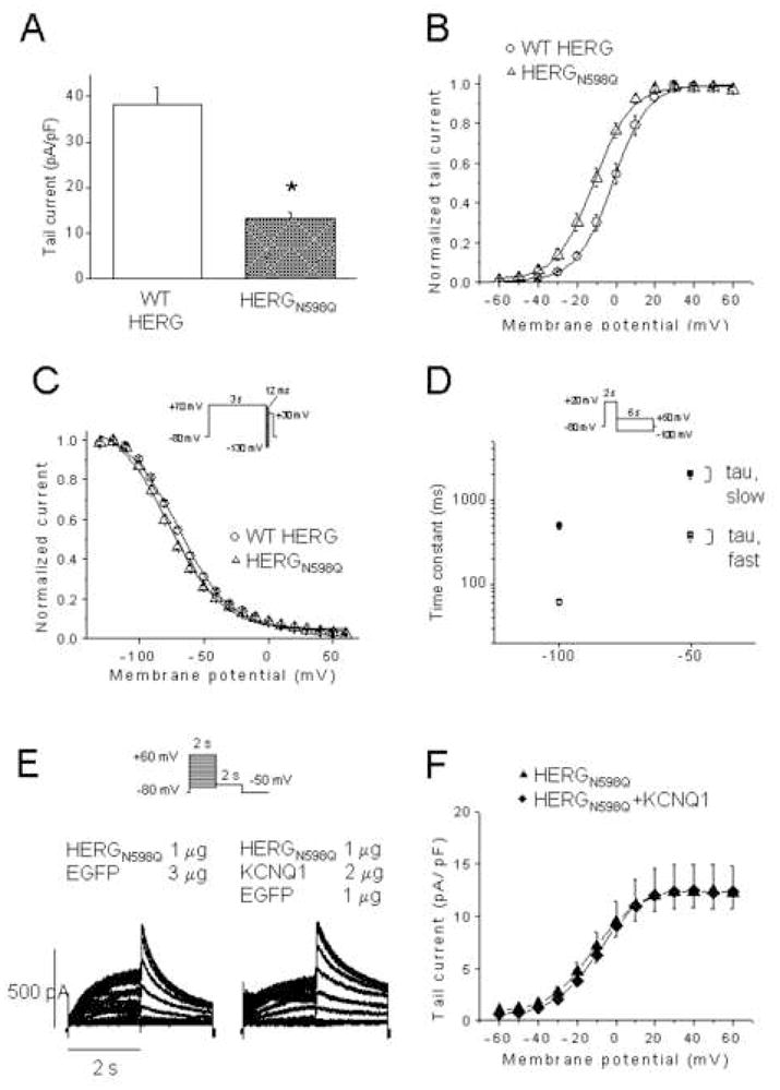
Figure 6. HERGN598Q with reduced membrane residence time, not rescued by KCNQ1.

(A) Tail current density measured at -50 mV after a test pulse to + 20 mV of WT HERG (n = 17) and HERGN598Q (n=19). (B), Normalized I-V relationships for tail currents of WT HERG (n = 12) and HERGN598Q (n=9). (C), Normalized steady-state inactivation curves of WT HERG (n = 8) and HERGN598Q (n=9). (D), Deactivation time constants of WT HERG (open and closed circles, n=11) and HERGN598Q (n=9). (E), Representative traces from CHO cells expressing HERGN598Q, and HERGN598Q+KCNQ1. Inset, voltage protocol. Current-voltage relationship of tail currents of HERGN598Q (n=15) and HERGN598Q + KCNQ1 (n=15). Dofetilide sensitive currents only are represented.
