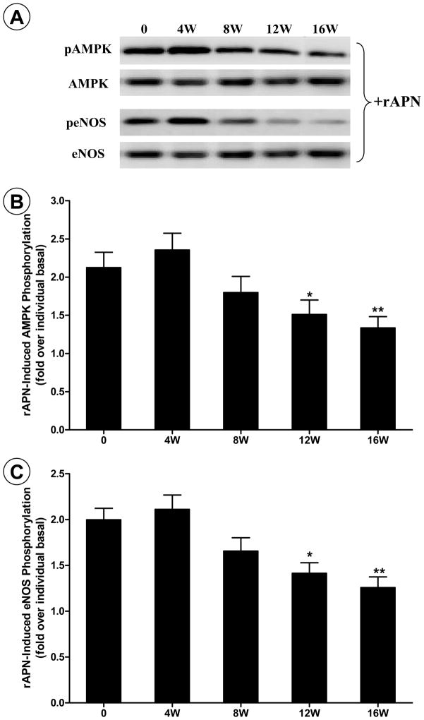
Figure 2.
Recombinant APN-induced AMPK and eNOS phosphorylation in aortic segments isolated from control rats, or rats fed high-fat diet (HFD) for 4 to 16 weeks. A: representative Western blot photos from aortic tissues stimulated with APN. B: ratio of pAMPK and AMPK with (solid bars) and without (open bars) APN stimulation; C: ratio of peNOS and eNOS with (solid bars) and without (open bars) APN stimulation. *P<0.05 and **P<0.01 vs. control rats. N=8–10 animals/group.

