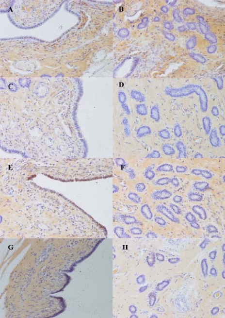
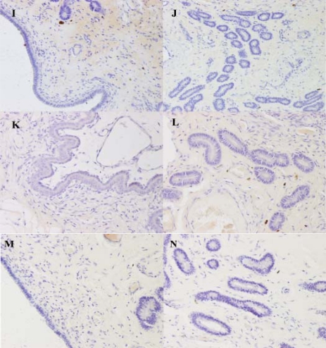
Figure 4.
Immunohistochemical localization of Muc1 in pig uterus. (A) and (B): Tissue from between attachment sites of a Day 13 pregnant sow. (C) and (D): At attachment sites of a Day 13 pregnant sow. (E) and (F): Between attachment sites of a Day 18 pregnant sow. (G) and (H): At attachment sites of a Day 18 pregnant sow. (I) and (J): Between attachment sites of a Day 24 pregnant sow. (K) and (L): At attachment sites of a Day 24 pregnant sow. (M) and (N): At attachment sites of a Day 13 pregnant sow, negative controls for localization (×200).


