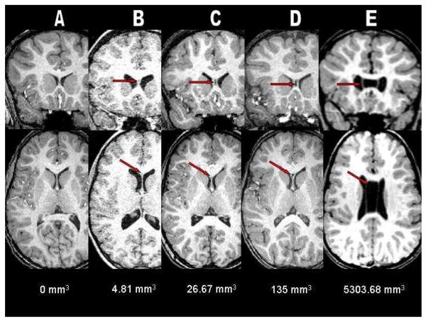
Figure 1.
A composite of coronal and axial T1-weighted anatomical magnetic resonance (MR) images illustrating an example from each of the five CSP classifications used in this study: A = None, B = Normal, C = Borderline, D = Abnormal, and E = Extreme. Note the complete nonfusion of the septal leaflets in E showing the presence and severe enlargement of cavum septum pellucidum/cavum verga in a patient with chromosome 22q11.2 deletion syndrome.

