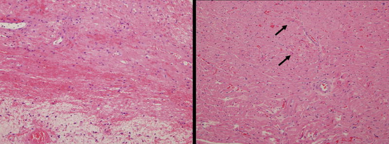
Figure 3.

Representative pathology of grafts. The left panel shows representative histology from groups A, B, C and E. The myocardium has marked coagulative necrosis with hemorrhage, edema and organization. There is minimal inflammation and most cells present are macrophages. These features are consistent with end stage rejection. The right panel shows hearts from Group D (those perfused with a solution of UW and Ad-NIS + 131I injection on POD 3). There are only focal areas of coagulative necrosis (arrows), but no cellular infiltrates. (Hematoxylin and eosin. Magnification × 200, both panels).
