
Figure 4.
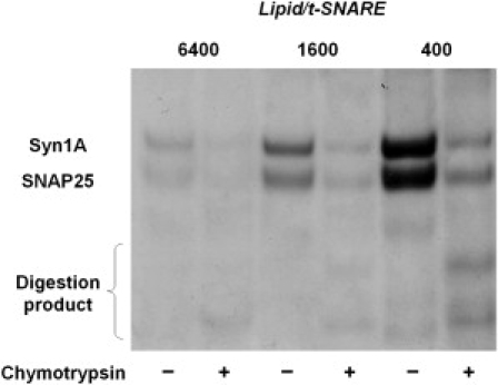
Orientation of SNAREs in the liposome membrane. In this example, proteoliposomes were prepared by direct incorporation of t-SNAREs into 50-nm liposomes. Upon addition of chymotrypsin, t-SNAREs facing outside were proteolyzed, whereas those facing the lumen of the liposomes were protected. Four t-SNARE liposomes with different lipid/protein ratios were exposed to chymotrypsin for 30 min at room temperature and then loaded onto an SDS-PAGE gel, juxtaposed with the same amount of corresponding untreated samples. The percentage of unprotected t-SNAREs, i.e., those exposing their cytosolic domain to the outside, was calculated by comparing the band intensity of the chymotrypsin-treated sample to that of the untreated sample. In this case, ∼75% of the t-SNAREs have their cytoplasmic domain oriented toward the outside of the liposomes. Statistics and results for other SNARE liposomes are displayed in Table 1, Table S1, and Table S2.