
Figure 1.
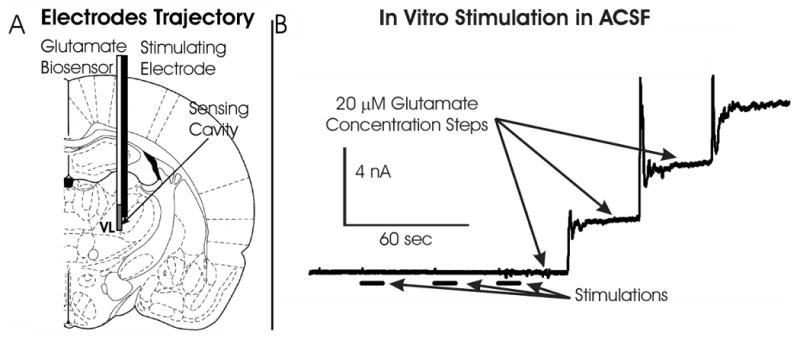
(A):Illustration showing the configuration of the electrical bipolar stimulating electrode and glutamate biosensor implanted in the rat thalamus to evoke and record glutamate release, respectively. (B): Oxidation current measured by the glutamate biosensor in artificial cerebrospinal fluid during 10 seconds monophasic stimulations (1 mA, 100 Hz, 100 μsec pulse width) and after 20 μM step increases in glutamate concentration.