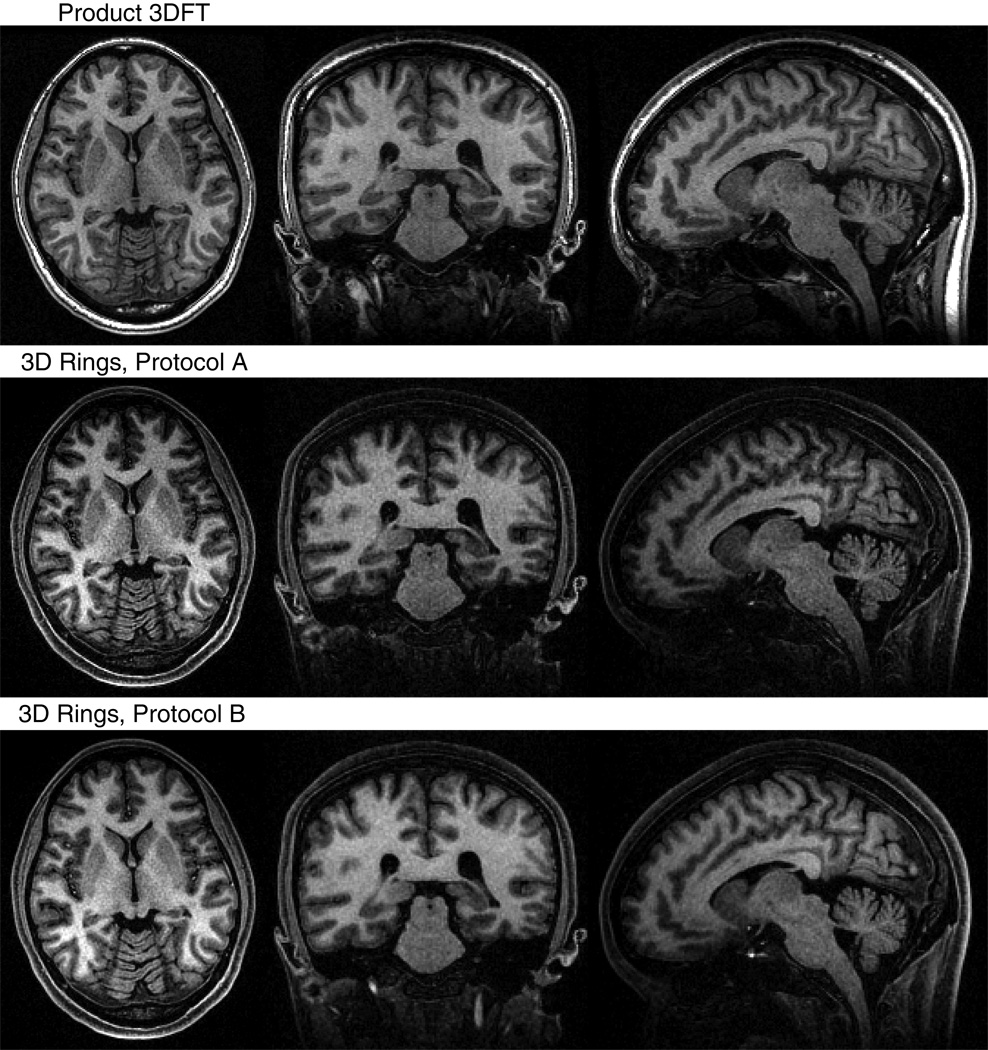
FIG. 8.
Comparison of 3DFT and 3D stack-of-rings scans for the same volume. In addition to protocols A and B, we also imaged the same brain volume using a product 3DFT IR-SPGR sequence. Only the reconstructed water images were used for comparison for the 3D stack-of-rings scans. Representative axial, coronal, and sagittal slices of the same position are displayed here. All scans show good WM/GM contrast. ROIs were localized in homogeneous areas of white matter and regions of cortical gray matter and the caudate nucleus for quantitative analysis. The 3D stack-of-rings scan using protocol A required roughly half the scan time of the 3DFT sequence, while achieving higher CNR and higher contrast efficiency. Protocol B required a slightly longer scan time than protocol A, but was still faster than the product sequence and achieved the highest SNR and CNR levels of the three. The actual measured values are listed in Table 1.

