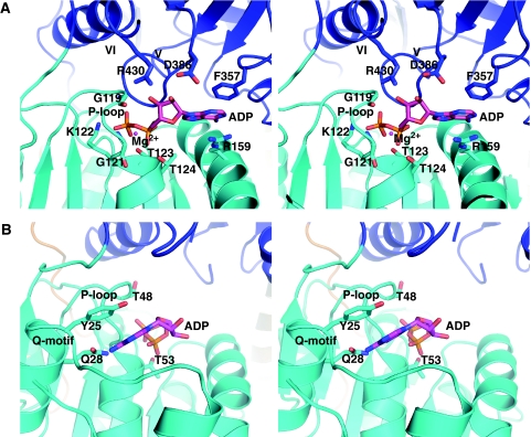
Figure 2.
Detailed comparison of the ATPase sites of Prp43p and the Hel308 homologue Hjm. The RecA1 and RecA2 domains are coloured as in Figure 1. Residues that directly interact with the ADP molecule (magenta sticks) are shown as sticks and labelled. The conserved sequence motifs are also indicated. (A) Close-up stereo view of the Prp43p ADP-binding site. The magnesium ion is pictured as a magenta sphere. (B) Close-up stereo view of the Hel308 homologue Hjm ADP-binding site (PDB 2ZJ5; Oyama et al, 2009) in the same orientation as in (A).

