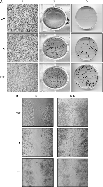
Figure 2.
During the focus-formation assay, OVCAR-3A and WT cells grew at a high density and formed numerous foci (A1). Anchorage-independent growth assays showing the development of clones in OVCAR-3A and LTE cells (A2). Plating efficiency. (A3) Migration assay, when ‘scratch’ wounds were created by scraping with a sterile pipette tip to make a gap. Only OVCAR-3LTE cells were not capable of settling the created ‘scratch’ wounds (B).

