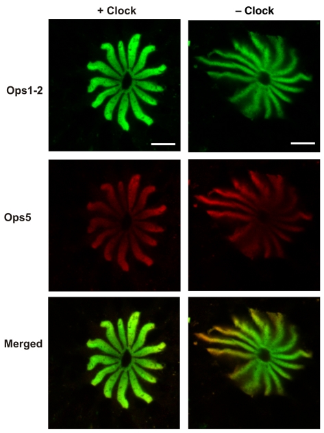
Fig. 10.
Ops1-2-immunoreactivity (Ops1-2-ir) and Ops5-ir in the R-segments and proximal A-segments of photoreceptors in frozen sections of lateral eyes (LEs) from a single animal fixed at night in the dark about 20 h after sunrise. The optic nerve to one of the LEs was cut to eliminate clock input (– Clock) at least 10 days before the experiment. The optic nerve to the other LE remained intact (+ Clock). Shown are images of sequential scans obtained from a single optical section and their merged images (Ops1-2, green; Ops5, red). Sections were immunostained at the same time, and images were collected in a single session using identical confocal settings. At night, Ops1-2 and Ops5 are highly localized to the rays of the rhabdom with little opsin-ir debris in the R-lobe or proximal A-lobe. This and other images suggest the ratio of rhabdomeral Ops5-ir to Ops1-2-ir is higher in LEs with cut lateral optic nerves (– Clock). Scale bars, 10 μm.

