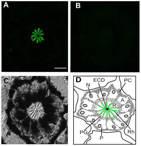
Fig. 5.
(A,B) Images of single optical sections of ommatidia from frozen sections of the same lateral eye (LE) fixed 20 h after sunrise in the dark. A and B were collected with identical confocal settings. (A) Ommatidium from a section immunostained with mAbOps5 (1:1000) that had been preincubated with a strip of nitrocellulose without antigen. Strong Ops5-immunoreactivity (Ops5-ir) is detected over the rays of the rhabdom. (B) Ommatidium from a section immunostained with mAbOps5 (1:1000) that had been preincubated with a strip of nitrocellulose to which ∼100 μg of antigen had been blotted. No Ops5-ir is detected. (C) Transmitted light image of the ommatidium shown in B. (D) Diagram of a cross section through one LE ommatidium, showing 12 photoreceptor cell bodies (P) with their arhabdomeral (A) and rhabdomeral (R) segments, rhabdom (Rh, in green), nucleus (N) and pigment granules (PG). The photoreceptors are surrounded by pigment cells (PC). In the center of the ommatidium is the dendrite of the eccentric cells (ECD), which is electrically coupled to the photoreceptors. Scale bar, 25 μm.

