Fig. 6.
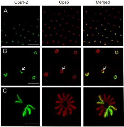
Ops1-2-immunoreactivity (Ops1-2-ir) and Ops5-ir in a frozen section of a lateral eye (LE) fixed 20 h after sunrise in the dark. Shown are images of a single optical section obtained with sequential scans of each fluorophore and their merged images. Ops1-2 (green); Ops5 (red). (A) A field of ommatidia in which all retinular cells are immunoreactive for Ops5 but only some are immunoreactive for Ops1-2. Many retinular cells in the upper left lack Ops1-2-ir. Scale bar, 300 μm. (B) Ommatidia from the field shown in A. In one ommatidium (lower right), all rhabdomeres are double labeled for Ops5 and Ops1-2; in the remaining three ommatidia, only some rhabdomeres are double labeled. The arrow points to the ommatidium shown in C. Scale bar, 100 μm. (C) All 11 rhabdomeral rays show Ops5-ir. One complete ray and half of four other rays are double labeled for Ops5 and Ops 1-2. Scale bar, 20 μm.

