Fig. 8.
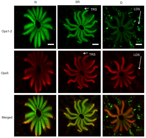
Ops1-2-immunoreactivity (Ops1-2-ir) and Ops5-ir in the R-segment and proximal A-segment of retinular cells in frozen sections of LEs fixed at different times of the day under natural illumination: during the night (N), between 18 and 20 h after sunrise, at sunrise (SR) and during the day (D), at ∼10 h after sunrise. Shown for each time point are images of sequential scans of a single optical section and their merged images (Ops1-2, green; Ops5, red). Sections were immunostained at the same time, and images were collected in a single session using identical confocal settings. At night, Ops1-2- and Ops5-ir are highly localized to the rays of the rhabdom, with little Ops-ir debris in the R-lobe or proximal A-lobe. At sunrise, levels of rhabdomeral Ops1-2- and Ops5-ir are similar to those seen during the night, but extra-rhabdomeral Ops1-2-ir and, to a lesser extent, Ops5-ir membranous debris, produced by transient rhabdom shedding (TRS), is detected in the R-segment and proximal A-segment. Later during the day (D), rhabdomeral Ops1-2-ir appears reduced compared with that observed in night-time and sunrise eyes, while intensely Ops1-2-ir extra-rhabdomeral membranous debris, produced by light-driven shedding (LDS), is detected in the R- and A-segments. By contrast, rhabdomeral Ops5-ir does not appear reduced in daytime eyes compared with eyes fixed during the night and at sunrise, although some extra-rhabdomeral Ops5-ir membranous debris is detected. At least some Ops5-ir co-localizes with Ops1-2-ir debris that is known to be in endosomes destined for degradation. Scale bars, 10 μm.

