
Figure 2.
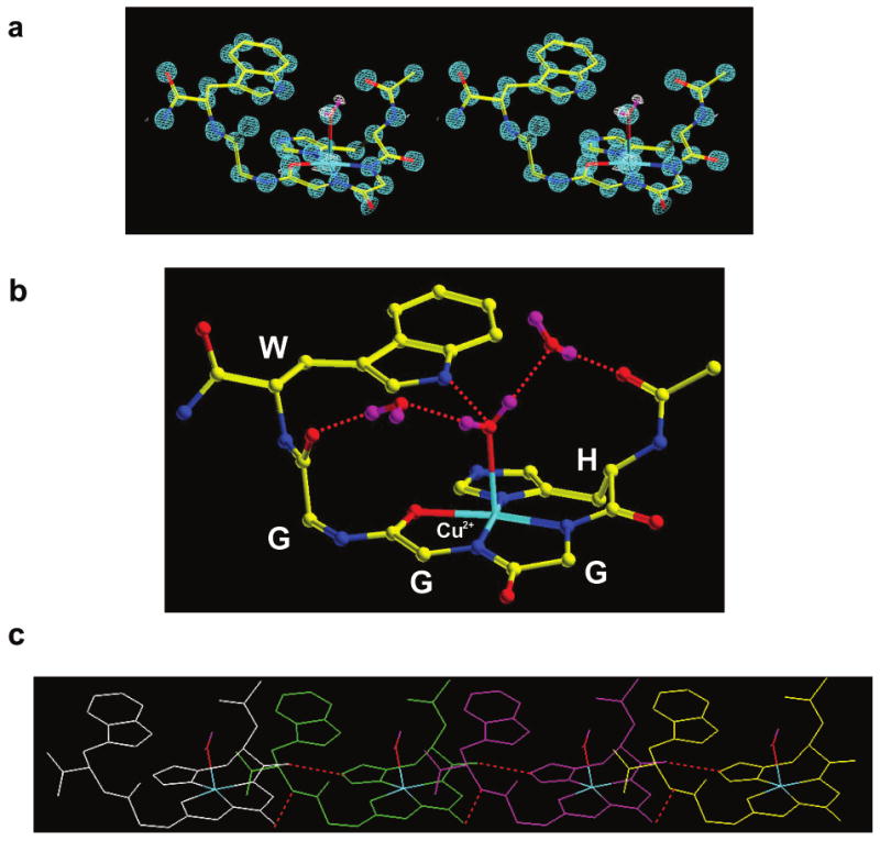
Crystal structure (0.7 Å resolution) of the HGGGW segment in complex with Cu2+. (a) A stereo representation of the electron density contoured at 2 σ is shown in blue. A difference map (white) reveals hydrogen density for the axially bound water. (b) This molecular representation shows how copper coordination is from the histidine imidazole and deprotonated amides from the next two glycines. In addition, the NH of the indole is within hydrogen bonding distance to the oxygen of the axial water. Two additional intramolecular ordered water molecules are also shown. (c) The red dashed lines show intermolecular hydrogen bond contacts identified in the crystal. These four copies of the copper binding units suggest a possible way in which the full octarepeat domain orders.