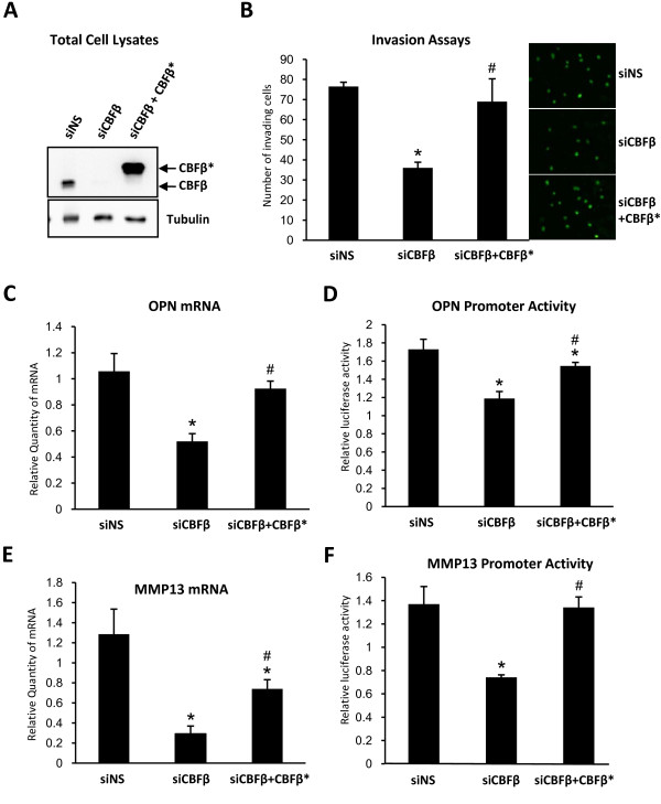
Figure 5.
Re-expression of CBFβ restores invasion capacity and OPN and MMP-13 expression. (A) Western blots showing re-expression of CBFβ after siRNA knockdown. The upper panel shows total cell lysates after immunodetection with an anti-CBFβ antibody. (B) Re-expression of CBFβ restores invasive capacity. MDA-MB-231 cells were transfected with siCBFβ or siNS. siCBFβ treated cells were transfected with the Flag-CBFβ-HA expression plasmid (CBFβ*). Cells that migrated through Matrigel were stained and counted in random fields. Data are presented as mean ± standard deviation (S.D.) (n = 3). * indicates p < 0.05 compared to siNS by analysis of variance; # indicates p < 0.05 compared to siCBFβ by analysis of variance. (C) Re-expression of CBFβ restores OPN gene expression. MDA-MB-231 cells transfected with siCBFβ or siNS followed by transfection with the CBFβ* expression plasmid. Data were analyzed as in (B). (D) Re-expression of CBFβ stimulates the OPN gene promoter. MDA-MB-231 cells transfected with siCBFβ or siNS were transfected with a plasmid containing the WT OPN promoter or a mutant OPN promoter containing mutated Runx2 sites. Cells were subsequently transfected with the CBFβ* expression plasmid. Reporter activity was obtained by comparison of the WT and mutant promoter activities. Data were analyzed as in (B). (E) MMP-13 gene expression is restored by re-expression of CBFβ. The experiment was performed as described in (C). (F) Re-expression of CBFβ stimulates the MMP-13 gene promoter. The experiment was performed as described in (D) using reporter plasmids containing the WT human MMP-13 promoter or a mutant MMP-13 promoter containing mutated Runx2 sites.

