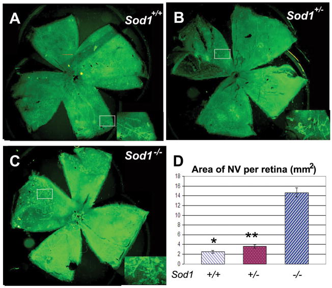
Figure 2. Mice deficient in superoxide dismutase 1 (Sod1−/−) show excessive amounts of ischemia-induced retinal neovascularization compared to Sod1+/− or Sod1+/+ mice.
Litters containing Sod1−/−, Sod1+/−, and Sod1+/+ pups were placed in 75% oxygen at postnatal day (P) 7, returned to room air at P12, and euthanized at P17. Retinal neovascularization on the surface of the retina was visualized by staining for PECAM-1 as described in Sod1+/+ (A) and Sod1+/− mice (B), but in comparison, Sod1−/− mice appeared to have substantially more neovascularization (C). Insets show a high magnification view of the retinal neovascularization present within the box in the whole mounts. Measurement of the area of neovascularization per retina by image analysis with the investigator masked with respect to genotype showed a marked increase in the mean (± SEM) area of neovascularization in Sod1−/− mice (n=7) compared to Sod1+/+ (n=10) and Sod1+/− (n=8) mice (D).
*p=0.0005; **p=0.0005 by ANOVA with Dunnett’s correction for multiple comparisons.

