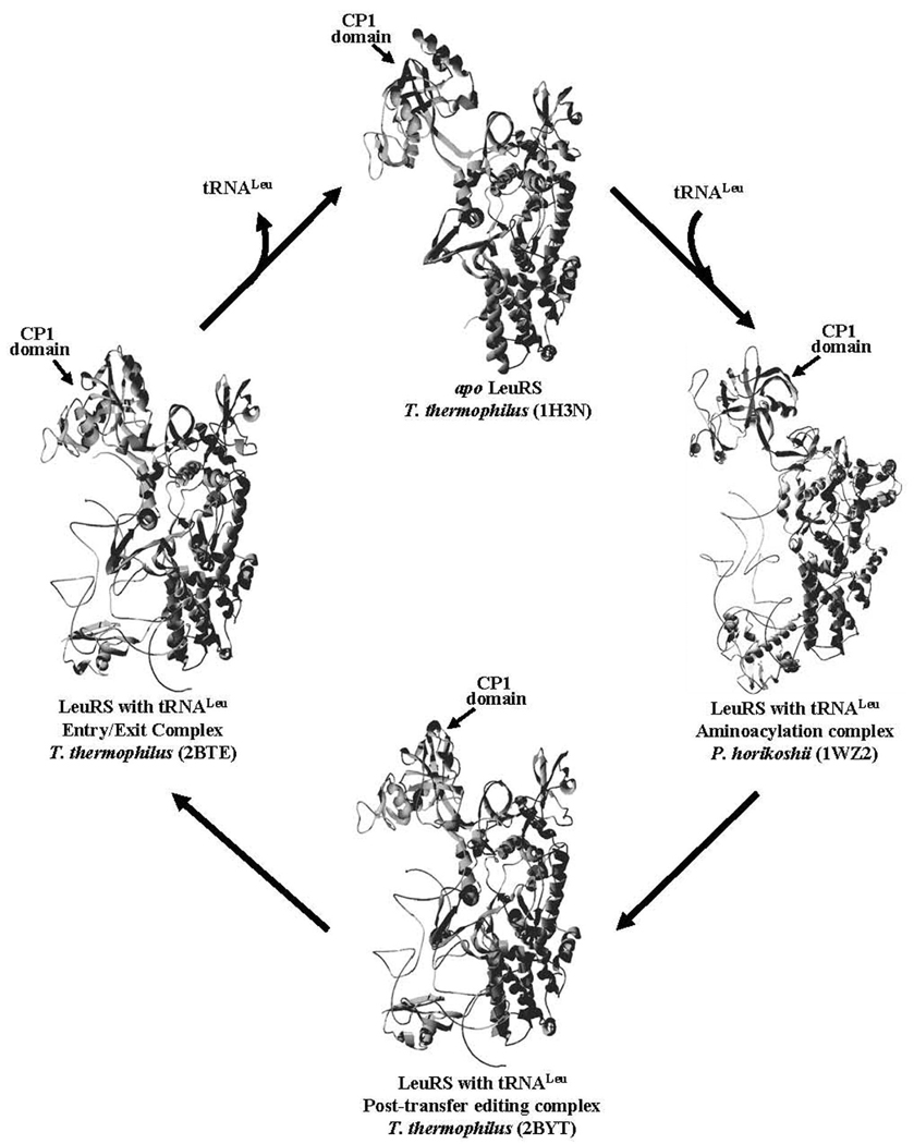
Figure 1.
Tertiary Structures of LeuRS. The enzyme cycle of LeuRS shows the apo X-ray crystal structure (top, PDB code: 1H3N), aminoacylation co-crystal structure complex with tRNA (right, PDB code: 1WZ2), and the post-transfer editing co-crystal complex (bottom, PDB code: 2BYT). The exit complex, which has also been proposed to be an entry complex for tRNA binding (29), is shown at the left (PDB code: 2BTE). The LeuRS protein structure is displayed as a ribbon, while the bound tRNA is indicated as a distinct line. The CP1 domain is oriented at the top left of each structure as indicated by marked arrow.

