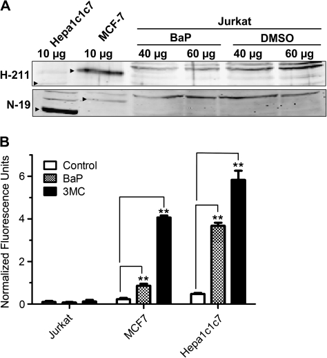
FIG. 1.
AhR expression and CYP1A1 activity in Jurkat cells. (A) Western blot analysis showing that AhR is not detected in BaP- and DMSO-treated Jurkat cells. Jurkat cells were treated with 2.5μM BaP or DMSO vehicle for 48 h. Different amounts of WCE from BaP- or DMSO-treated Jurkat (40 and 60 μg), MCF-7 (10 μg), and Hepa1c1c7 (10 μg) cells were used to detect the AhR protein using AhR-specific antibodies H-211 (upper panel) and N-19 (lower panel). Arrowheads indicate the human (MCF-7) and mouse AhR (Hepa1c1c7). This Western has been repeated at least once. (B) CYP1A1 activity of Jurkat, MCF7, and Hep1c1c7 cells determined by EROD assay. Cells were treated with DMSO vehicle, 2.5μM BaP, or 1μM 3MC for 5 h at 37°C, followed by EROD assay. The error bars represent ±SD of triplicate samples (n = 3, ** p ≤ 0.005). Normalized fluorescence units were calculated by subtracting each fluorescence number with the number obtained from the same treatment without cells.

