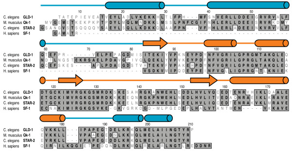
Figure 1.
STAR domain sequence alignment between C. elegans GLD-1 and STAR-2, mouse Qk-1 and human SF-1. Positions with identical or similar amino acid residues are boxed in dark gray and differences are unboxed in white. Secondary structure elements above the sequence alignment are from the crystal structure of the GLD-1 Qua1 domain [15] (PDB ID 3K6T) and the solution structure of the SF-1 KH-Qua2 domains [11] (PDB ID 1K1G) with α-helices and β-strands represented by cylinders and arrows, respectively. Qua1 and Qua2 secondary structure domain boundaries are colored in blue and the central KH domain is colored in orange.

