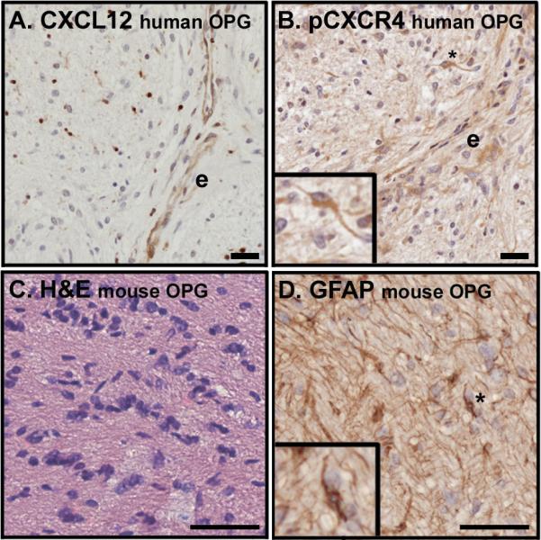
Figure 1. Optic nerve gliomas in humans and mice.

(A) An optic nerve glioma specimen from a patient with NF1 demonstrates mild hypercellularity and CXCL12 expression in the endothelium of tumor-associated blood vessels (e) as well as in scattered infiltrating cells. (B) Serial section from the tumor presented in panel A stained for the presence of a ligand-induced phosphorylated form of CXCR4 (pCXCR4) reveals a high level of receptor activation in proximity to the CXCL12-expressing endothelium. Phosphorylated CXCR4 is present in a pilocytic cell (inset). (C) A hypercellular lesion with nuclear atypia is evident within the optic nerve of an OPG mouse with hematoxylin and eosin stain. (D) The tumor pictured in panel C contains GFAP-expressing cells with the elongated bipolar morphology characteristic of pilocytic cells. In all cases expression appears brown. Scale bars for A and B equal 20 microns and for C and D equal 50 microns.
