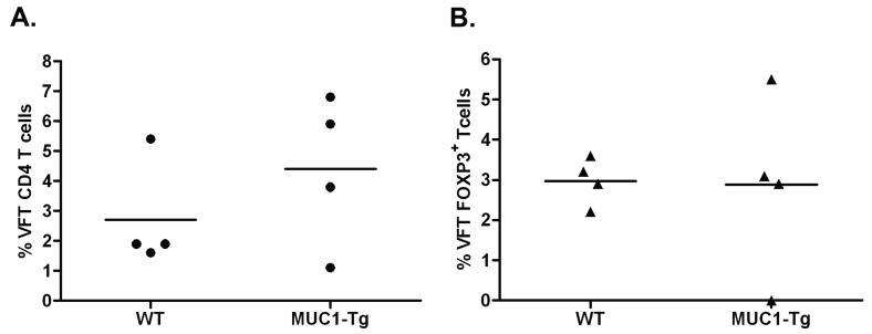
Figure 1. VFT precursors develop through the thymus and enter the periphery at equal levels in WT and MUC1-Tg mice.
Recipient mice (WT, n=4 and MUC1-Tg, n=4) were lethally irradiated prior to bone marrow transfer. Five weeks post VFT bone marrow transfer, the presence of mature donor VFT CD4 T cells in the spleens of recipient mice was assessed by flow cytometry. A) Percent of donor cells (Vα2+CD90.1+) in the CD3+CD4+ gated population of each recipient mouse. B) Intracellular Foxp3 expression in donor cells. Mean values are shown (—). These data are representative of two independent experiments.

