Figure 3.

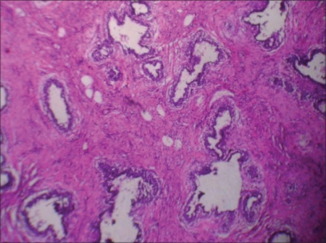
Histopathology (H and E Stain; Photomicrograph- 10 × 10) shows increased cellularity of Stromal and parenchymal component. no periductal concentrates of cells

Histopathology (H and E Stain; Photomicrograph- 10 × 10) shows increased cellularity of Stromal and parenchymal component. no periductal concentrates of cells