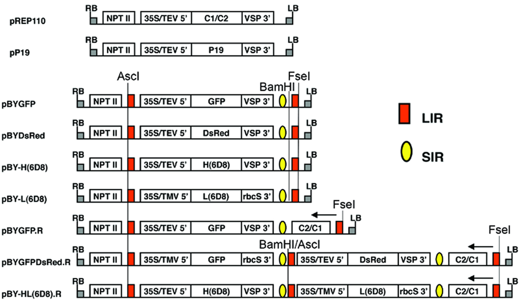
Figure 1.
Schematic representation of the T-DNA regions of the vectors used in this study. 35S/TEV 5’, CaMV 35S promoter with tobacco etch virus 5’ UTR; 35S/TMV 5’, CaMV 35S promoter with tobacco mosaic virus 5’ UTR; VSP 3’, soybean vspB gene 3’ element; rbcS 3’, pea rbcS gene 3’ element, NPT II, expression cassette encoding nptII gene for kanamycin resistance; LIR (red box), long intergenic region of BeYDV genome; SIR (yellow oval), short intergenic region of BeYDV genome; C1/C2 (pREP110) or C2/C1 (pBYGFP.R, pBYGFPDsRed.R, and pBY-HL(6D8).R), BeYDV ORFs C1 and C2, encoding replication initiation protein (Rep) and RepA; LB and RB, the left and right borders of the T-DNA region. Arrows at right-most LIR in pBYGFP.R, pBYGFPDsRed.R, and pBY-HL(6D8).R indicate direction of transcription, from the LIR promoter, of the complimentary sense genes C1/C2. In the dual replicon constructs pBYGFPDsRed.R and pBY-HL(6D8).R, the middle LIR functions in concert with the left-side LIR to release the left-side replicon, and it also functions in concert with the right-side LIR to release the right-side replicon. In pBYGFPDsRed.R and pBY-HL(6D8).R, BamHI/AscI marked at the middle LIR is the site of fusion of two replicons via the Klenow-filled BamHI site of the left-side replicon and the Klenow-filled AscI site of the right-side replicon.

