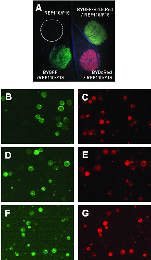
Figure 2.
Expression of GFP and/or DsRed replicons in plant leaf cells. A: N. benthamiana leaves infiltrated with Agrobacterium strain harboring expression vectors as indicated were examined and photographed at 5 days post infiltration (dpi) under a handheld UV lamp as described in Materials and Methods. Co-infiltration of a replicon (pBYGFP or pBYDsRed) with pREP110 allows replication of the DNA lying between the LIR regions (Fig. 1). Co-infiltration with pP19 suppresses post-transcriptional gene silencing. B – G: Fluorescence microscopy of mesophyll protoplasts expressing GFP and/or DsRed replicons. Mesophyll protoplasts were prepared from infiltrated leaf tissue at 5 dpi and subsequently viewed under a Nikon inverted microscope with both GFP and DsRed filters. B and C: mixture of protoplasts from leaves infiltrated separately with BYGFP/REP110/P19 or BYDsRed/REP110/P19. D and E: protoplasts from leaves infiltrated with BYGFP/BYDsRed/REP110/P19. F and G: protoplasts from leaves infiltrated with BYGFPDsRed.R. Panels B, D and F are viewed with a GFP filter; and C, E and G are viewed with a DsRed filter. Almost all cells in panels D, E, F, and G show both green and red fluorescence, indicating co-expression of GFP and DsRed.

