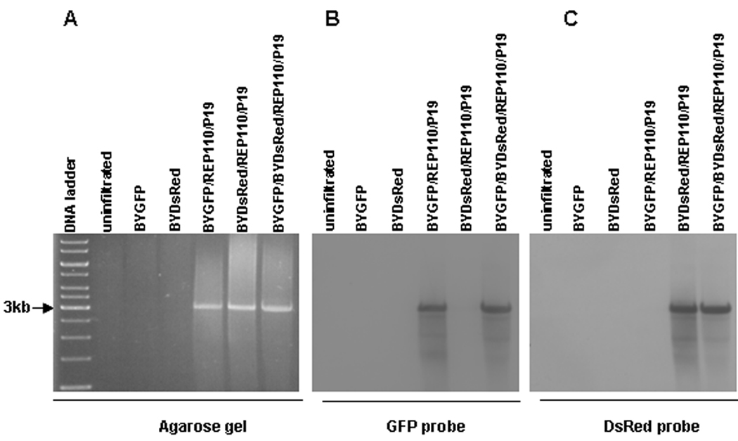
Figure 3.
Southern blot showing non-competing formation of two replicons. Leaf DNA was extracted at 5dpi from leaves infiltrated with the indicated vector combinations, digested with XhoI, and run on a 1% agarose gel. The resulting gel was stained with ethidium bromide (A), blotted onto membrane, and detected with a GFP probe (B). The same membrane was subsequently stripped and re-hybridized with a DsRed probe (C). The preparation of GFP- and DsRed-specific probes is described in Materials and Methods. Arrow at left (3kb) marks the size of the major DNA species observed on both stained gel and Southern blots. Both GFP and DsRed replicons are ~3kbp, but note that the GFP probe does not bind to the DsRed replicon (B), and vice versa (C).

