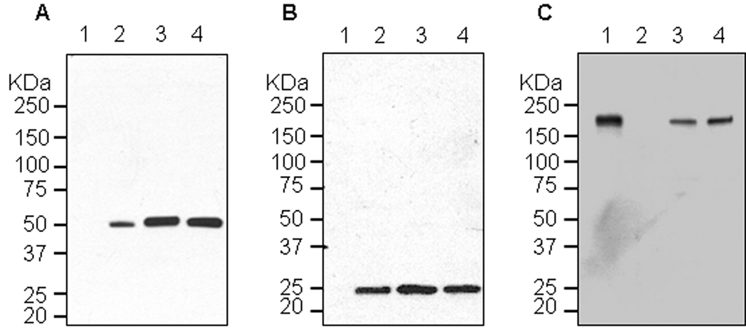
Figure 5.
Western blot analysis of plant-derived 6D8. Protein samples were separated on a 4–12% SDS-PAGE gradient gel under denaturing and reducing condition (A and B) or under non-reducing condition (C) and blotted onto a PVDF membrane. The membrane was incubated with a goat anti-human gamma chain antibody or goat anti-human kappa chain antibody to detect heavy chain (A) or light chain (B and C). Lane 1, Protein samples extracted from un-infiltrated leaves (A and B), or human IgG as a reference standard (C); lane 2, human IgG as a reference standard (A and B), or Protein samples from un-infiltrated leaves (C); lane 3, Protein sample extracted from the leaves co-infiltrated with separate replicons for light chain (pBY-L(6D8)) and heavy chain (pBY-H(6D8)) ; lane 4, Protein extracted from the single vector pBY-HL(6D8).R infiltrated leaves.

