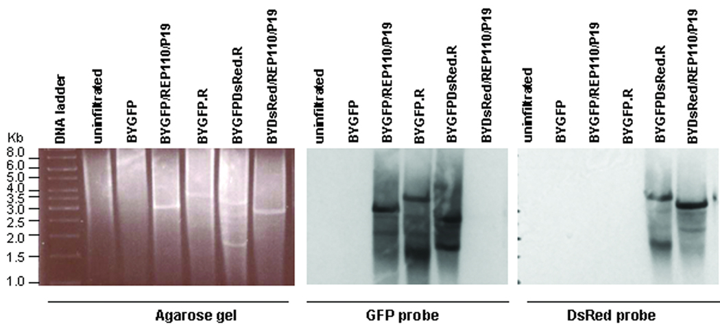
Figure 6.
Formation of two separate replicons by a single vector. DNA from leaves infiltrated with various vector combinations as indicated was digested with XhoI, run on 1% agarose gel and stained with ethidium bromide (A), blotted onto membrane, and detected with a GFP probe (B) or a DsRed probe (C). The preparation of GFP- and DsRed-specific probes is described in Materials and Methods. Numbers at left indicate sizes in kbp of DNA molecular weight markers. Note that pBYGFP produced a replicon of ~3kbp (consistent with data in Fig. 3), while the replicon from pBYGFP.R is ~4kbp due to the presence of the C1/C2 gene. The dual replicon construct pBYGFPDsRed.R produces a GFP replicon at ~3kbp and a DsRed replicon containing the C1C2 gene (Fig. 1) at ~4kbp.

