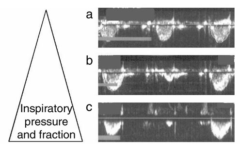
Figure 1.

Beat-to-beat changes in stroke volume: ultrasound Doppler records from three consecutive heart beats performed from the apex with the sample volume positioned in the left ventricular outflow tract, (a) at basal ventilator settings; (b) when inspiratory time was prolonged; (c) when inspiratory time as well as PIP was increased. Detailed information on ventilator settings for (a), (b) and (c) is presented in Table 1.
