
Figure 2.
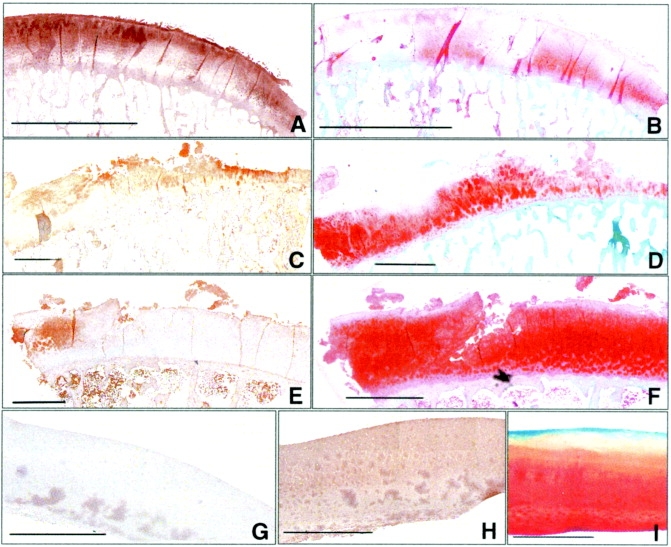
Binding of 1-11E to cartilage in patients with rheumatoid arthritis (RA) and osteoarthritis (OA). Antibody 1-11E diffusely stained RA cartilage in all layers (brown). A, Staining of the RA specimen in the superficial area and the middle zone was stronger than that in the deep zone. B, Staining of the RA specimen with Safranin O was weak and localized mainly in the deep zone (red). C, Staining of an OA cartilage sample with extensive erosions and marked surface damage, including the formation of fragments discrete from the parent cartilage, was strong in the most severely damaged area. D, Staining with 1-11E colocalized with an area of weak Safranin O staining in a parallel nonconsecutive OA cartilage section. E and F, Antibody 1-11E staining of cartilage from a patient with mild OA with typical fissuring of the surface of the upper cartilage appeared as a territorial “halo” around the chondrocytes (E), while staining with Safranin O was strong (F). G–I, Staining of the subchondral bone was not observed in any of the samples tested. No staining with 1-11E was detected in histologically normal cartilage (G), which also stained normally with a commercial anti–type II collagen monoclonal antibody (H) and with Safranin O (I). Bars in A and B = 5,000 μm; bars in C–H = 2,000 μm; bar in I = 500 μm.