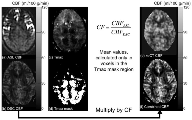
FIG. 1.

Demonstration of the method of determining the CF for combined ASL and DSC CBF maps. a: ASL- and (b) DSC-derived MRI CBF maps in a 42-year-old man with bilateral moyamoya disease. There is marked arterial transit artifact in the bilateral anterior circulation seen on the ASL CBF maps, while the posterior circulation is unaffected. Using information from the Tmax map (c) acquired during the DSC study, a thresholded Tmax mask (d) can be created to include only voxels with Tmax shorter than a prespecified threshold (in this example, Tmaxthresh = 3 sec). The CF is the ratio of the ASL-measured CBF and the DSC-measured CBF calculated only in the Tmax masked regions. The DSC-derived CBF map is then multiplied by the CF to create the combined map (f), which more closely compares with the xeCT CBF map (e) in magnitude while more accurately depicting true CBF in the affected regions.
