
Figure 1. “On” and “Off” states for ITAM Phosphorylation.
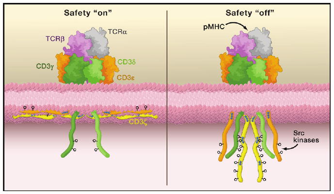
Shown on top are the subunits comprising the extracellular portion of the T cell receptor-CD3 complex: TCRα (gray), TCRβ (purple), CD3γ (dark green), CD3δ (light green), and CD3ε (orange). The cytoplasmic domains, including CD3ζ (yellow), are shown on the bottom along with the inner leaflet of the plasma membrane. The blue dots indicate stretches of basic residues that mediate interactions with the acidic inner leaf of the plasma membrane, while the phenyl rings indicate the critical tyrosines within the ITAM motifs. On the left, the TCR triggering safety is “on” and the ITAM motifs of the CD3ε and CD3ζ cytoplasmic domains are depicted as partially embedded within the plasma membrane with the tyrosines buried in the bilayer. On the right, the safety is “off” due to ligand binding (here as an antigenic peptide embedded in a major histocompatibility complex molecule [pMHC]); this may also occur spontaneously. In this “off” state, the critical tyrosine residues in the ITAM motifs become accessible to cytoplasmic Src kinases and become phosphorylated.