
Figure 3.
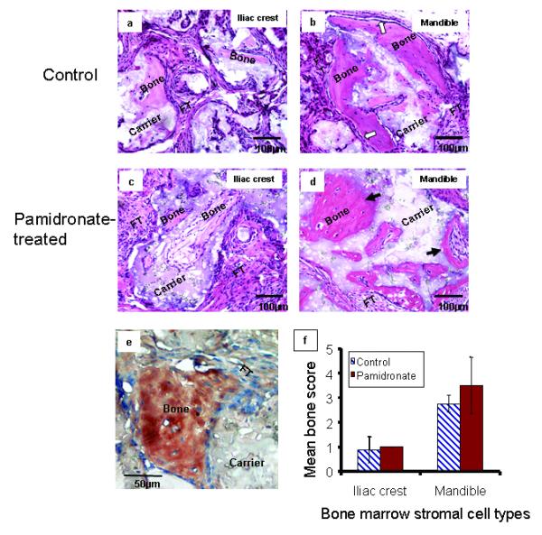
In vivo bone formation by pamidronate-treated mandible and iliac crest BMSCs Hematoxylin/eosin-stained sections of bone formed by transplanted control (a and b) and pamidronate-treated (c and d) BMSCs demonstrated that mandible BMSCs (b and d) formed more histologically observable bone than iliac crest BMSCs (a and c) as previously reported (Akintoye et al., 2006). Note distinct osteoblastic rimming indicative of bone lining cells in mandible control samples (white arrows, b). After pre-treatment with pamidronate followed by transplantation, mandible BMSCs formed structurally less-organized osteoid-like bone with fewer cells and loss of osteoblastic rimming (dark arrows, d) but iliac crest BMSCs formed a more fibrocellular bone matrix with cell-lined surface. Representative immunohistochemical staining with rabbit anti-human osteopontin (mandible control shown) confirms that bone formed by transplanted BMSCs were of human origin and not mice (e). Semi-quantitative analysis of in vivo bone showed that BMSCs pre-treated with pamidronate recovered from effects of pamidronate to form appreciable bone although not significantly higher than their respective controls (f). Higher quantitative bone by both control and pamidronate-treated mandible BMSCs further confirm their better ectopic bone-forming capacity relative to iliac crest BMSCs. (Data presented are representative histology sections and mean of n = 6 subjects).