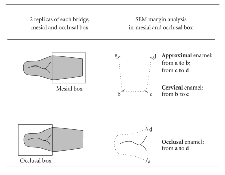
Table 2.
Scheme of the quantitative margin analysis in the Scanning Electron Microscope. Two replicas were obtained from each cantilever FPD; one from the mesial box and the other one from the occlusal box. For the quantitative margin analysis, the enamel was divided into three segments: interproximal (segments a-b and c-d), cervical (segment b-c) and occlusal (segment a-d). All segments together constituted the total margin length.

|
