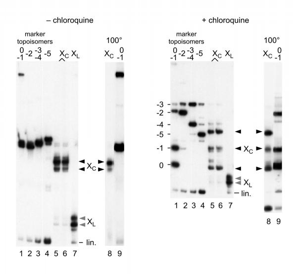
Figure 2.
Circularization of Form X. In these experiments a 258 bp linear fragment containing the same 60 bp tract of poly(CA) · poly(TG) as above was used. Linear Form X (bands labelled XL lane 7, greyed arrowheads) was incubated in the presence of DNA ligase, and the circular forms obtained (bands labelled Xc lanes 5 and 6, black arrowheads) were analyzed by electrophoresis on polyacrylamide gels in the absence (left panel) or in the presence (right panel) of 20 μM chloroquine. A series of marker topoisomers was prepared by ligation of the regular 258 bp linear fragment in the presence of variable amounts of ethidium bromide [10], yielding a series of topoisomers containing increasing numbers of negative supercoils (lanes 1-4), with up to 5 negative superturns for the most supercoiled topoisomer. In lanes 8 and 9, circularized Form X and topoisomers 0 and -1 were analyzed after incubation for 5 min. at 100°C. Note that circularized Form X does not migrate like any of the topoisomer markers.

