Figure 3.
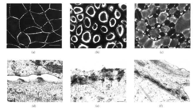
Desmosomes of MDCK cells can be calcium-dependent or calcium-independent. (a) Desmosomes of MDCK cells in confluent culture stained with monoclonal antibody to desmoplakin. Note that the desmosomes are located at the cell peripheries and that the staining is generally punctate. (b) A monolayer that has been cultured at confluent density for 24 hours in SM and then treated with LCM-EGTA for 90 minutes, showing loss of intercellular contact and of desmosomal staining from the cell peripheries. This is indicative of calcium-dependent desmosomes. (c) A 6-day-confluent monolayer treated with LCM-EGTA for 90 minutes, showing partial loss of intercellular contact but persistence of joining processes with intense desmosomal staining (e.g., arrow). This is indicative of calcium-desmosomes. Bar, 20 m. (d) When subconfluent or 1-day-confluent cells are treated with LCM-EGTA adhesion is lost and desmosomal halves separate. The half desmosomes are then internalised ((e), arrows). (f) By contrast when 6-day-confluent cells are treated with LCM-EGTA desmosomes remain adherent and accumulate at the cell surface between interconnecting cell processes. Bars: 0.3 m in (d) and (f); 0.4 m in (e) (a)–(c). Reproduced from the study by Wallis et al. in [37]. The isoform of protein kinase (c) is involved in signalling the response of desmosomes to wounding in cultured epithelial cells. (d)–(f) Reproduced from the study by Mattey and Garrod in [50].

