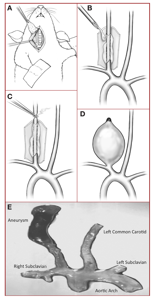
FIGURE 1.
A, drawing illustrating microsurgical exposure of the murine right common carotid artery (RCCA). B, drawing showing latex cuff placed around the RCCA, which is then bathed with porcine pancreatic elastase solution for 20 minutes. C, drawing showing distal occlusion of the RCCA. D, drawing showing a saccular aneurysm that has formed during the 3 weeks postinjury. E, photograph of a typical murine aneurysm (original magnification, ×1).

