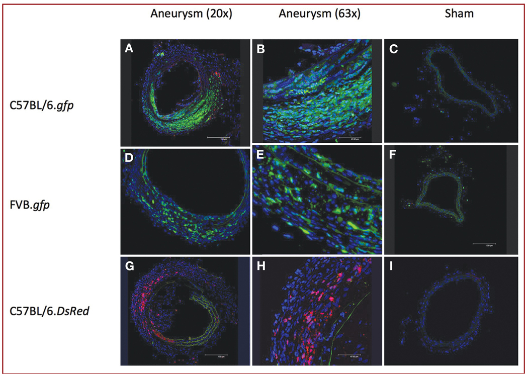
FIGURE 3.
Immunohistochemical staining showing abundant bone marrow–derived cells in the walls of aneurysms (original magnification, ×20 [A, D, and G] and ×63 [B, E, and H]), but not in sham-operated RCCAs (original magnification, ×20 [C, F, and I]) of C57BL/6.gfp (A–C), FVB.gfp (D–F), and C57BL/6.DsRed (G–I) radiation chimeric mice. Blue, 4′,6-diamidino-2-phenylindole (DAPI); green, gfp cells; red, DsRed cells.

