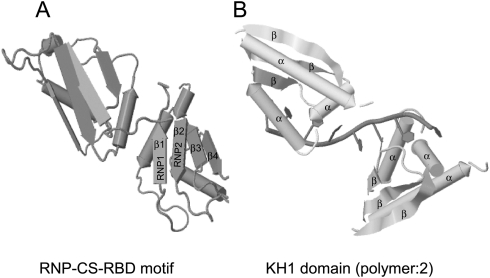
FIGURE 1.
Structure of hnRNP proteins. (A) The RNP-CS-RBD motif has a βαββαβ structure. The octameric RNP1 and hexameric RNP2 sequences are juxtaposed on β3 and β1, respectively. The structure shown is of multiple isomorphous replacement (MIR) of two RBDs of human hnRNP A1 at 1.75 Å (PDB ID: 1HA1) (Shamoo et al. 1997). (B) The KH domain comprises a triple β-sheet platform supporting three α-helical segments. Structure shown is of X-ray crystallography (resolution 3 Å) of KH1 domain (2-polymer) of human hnRNP E1 (PBD ID: 1ZTG) (Sidiqi et al. 2005). Both structures have been derived by the JMol algorithm.

