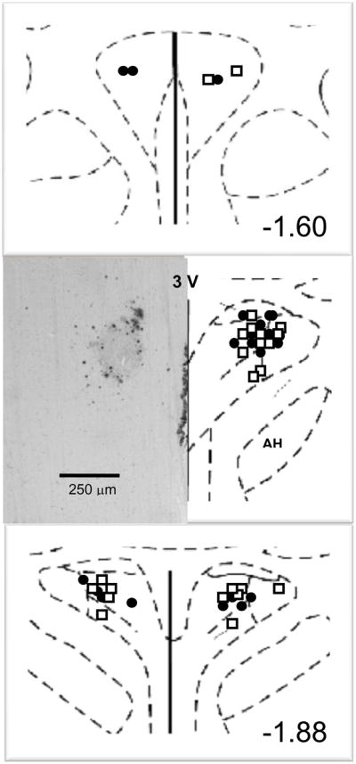
Figure 1. Microinjection site into paraventricular nucleus.

Typical microinjection site into paraventricular nucleus with β-galactosidase (left) and schematic diagram (right) of coronal section through the paraventricular nucleus are shown. The distance caudal to bregma indicated in the lower right corner is for the mature adult rat at the time of brain harvesting rather than the coordinates for microinjection used in the immature rat five weeks earlier to achieve the targeted area within the paraventricular nucleus. PVN, paraventricular nucleus; AH, anterior hypothalamus, 3V, third ventricle. •, injection sites for sham clipped groups; □, injection sites for two-kidney one-clip groups
