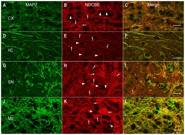
Fig. 8.
Indirect immunofluorescence of NDCBE in mouse brain sections. The sections were double-stained with a monoclonal antibody against the neuronal marker MAP2 (green) and the affinity-purified NDCBE antibody (red). (A–C) Cerebral cortex (CX). (D–F) Hippocampus (HC, the region between the CA1 region and dentate gyrus). (G–I) Substantia nigra (SN). (J–L) Medulla (Md). Arrowheads point to neuron cell bodies, and arrows point to processes. Scale bar: 20 μm.

