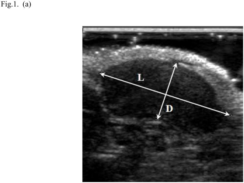
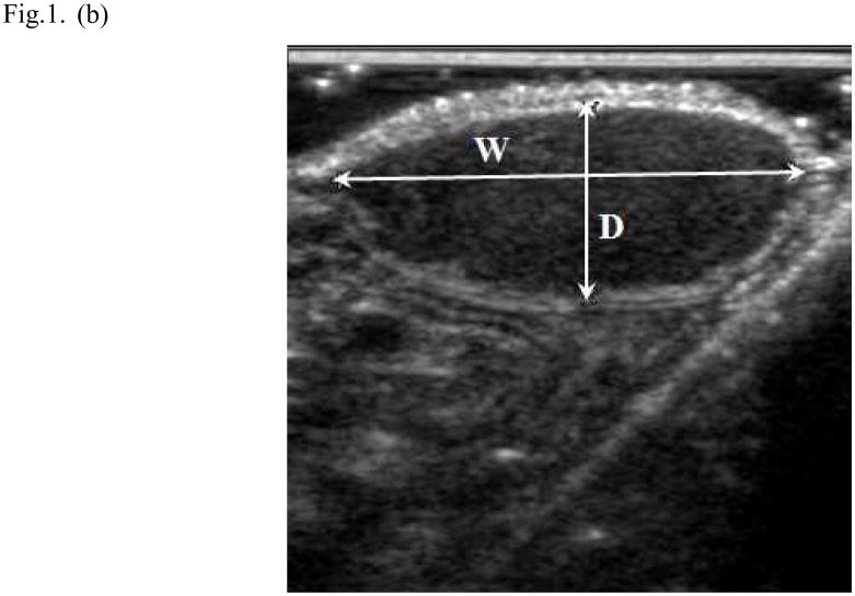
Fig. 1.
Ultrasound method for measuring tumor volume. In the longitudinal image (a) the length (L) of the tumor was 1.73cm and its depth (D) was 0.75cm; in the transverse image (b) the width (W) of the tumor was 1.65cm and its depth was 0.68cm. The volume (V) of the tumor was 1.02 mL was calculated (V = 0.5 LWD; where the depth measured in the two image planes was averaged).


