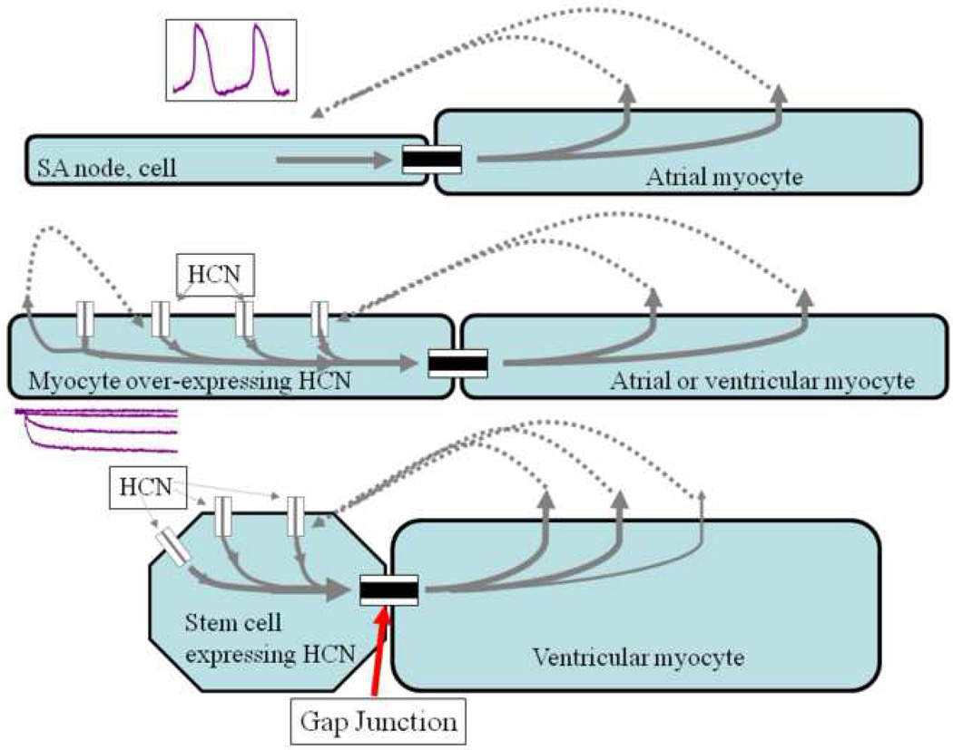
Figure 2.
Rationale for biological pacing. Top) Initiation of spontaneous rhythms by sinoatrial node cells. Action potentials (inset) are initiated via inward current flowing through transmembrane channels (see Figure 1). Current flowing via gap junctions to adjacent myocytes results in excitation and impulse propagation through the conducting system. Middle) Gene-based biological pacemaker implanted in atrium or ventricle. HCN channel genes are virally introduced into a group of myocytes (one shown), resulting in channel proteins incorporated in the myocyte membrane. When the membrane is hyperpolarized at the end of an action potential, the HCN channels open to induce inward current, which excites the myocyte to initiate an action potential that then propagates via gap junctions to neighboring cells. Bottom) Cell-based biological pacemaker shown implanted in ventricle. HCN channels are expressed in stem cells that are then implanted in myocardium, where they electrically couple to neighboring myocytes via gap junctions. When the stem cell membrane is hyperpolarized via current flow through the gap junctions from coupled myocytes, the HCN channels open to induce inward current, which travels through the gap junction to excite the coupled myocyte and initiate an action potential in the myocyte. In each depicted example, current spread requires the presence of gap junctions to electrically couple the cells (adapted with permission from [34]).

