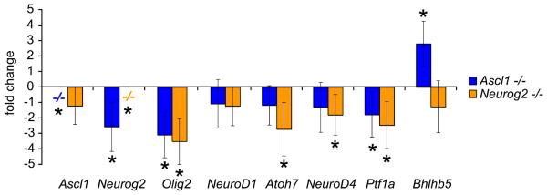
Figure 7. Ascl1 and Neuorg2 are upstream bHLH transcription factors.
QPCR was used to measure changes in other bHLH transcription factor family members expressed during retinal development due to loss of Ascl1 or Neurog2 (P0, see Figure 4, and Methods). Ascl1 was not expressed in the Ascl1−/− retina, and was not changed with loss of Neurog2 function. Likewise, Neurog2 was not expressed in the Neurog2−/− retina, however Neurog2 expression was decreased in the Ascl1−/− retina. Olig2 expression was decreased in both Ascl1 and Neurog2 knockout retina. NeuroD1 expression was not changed in either Ascl1 or Neurog2 knockout retina. Atoh7 and NeuroD4 expression were decreased in the Neurog2−/− retina, but not changed in the Ascl1−/− retina. Ptf1a expression was decreased in both Ascl1−/− and Neurog2−/− retina. Bhlhb5 expression was increased in the Ascl1−/− retina, but was not changed in the Neurog2−/− retina. These changes are consistent with the roles of Ascl1 in particular, as well as Neurog2, as upstream bHLH transcription factors expressed in progenitor cells that regulate a downstream proneural bHLH transcription factor cascade underlying the transition to differentiating neurons (Nelson et al., 2007a).

